
Calcinosis cutis in dogs is one of those skin problems that looks local but often reflects something deeper. Calcium salts can build up in the dermis and form gritty plaques, crusts, or hard nodules, and the real work is finding what pushed the skin to mineralize in the first place. In this article, I cover the signs to watch for, how veterinarians confirm the diagnosis, what treatment actually looks like, and how recovery usually unfolds.
Key points to know before you act
- Most canine cases are linked to excess corticosteroids, either from Cushing's disease or long-term steroid use.
- The lesions often feel gritty or hard and may show up on the neck, back, groin, armpits, or abdomen.
- Ulceration and chalky discharge can happen, especially if the area becomes inflamed or infected.
- Bloodwork helps look for the trigger, but skin biopsy is usually what confirms the diagnosis.
- Treatment works best when the underlying cause is controlled; topical DMSO, wound care, and surgery are add-ons, not substitutes.
- Improvement is often slow, and skin changes may take several months to clear even after the cause is addressed.
What calcinosis cutis means in dogs
When I look at this condition clinically, I think of it as a skin mineralization pattern, not a stand-alone skin disease. Calcium salts, often calcium phosphate, deposit in the skin and make the tissue feel firm, rough, or even sandpapery under the fingers.
There are two practical forms to keep in mind. The more familiar one is the generalized or steroid-associated form, which is commonly tied to Cushing's disease or prolonged glucocorticoid use. The other is a more localized form, often called calcinosis circumscripta, which tends to show up as a single nodule or a small cluster of nodules after trauma, repeated pressure, or irritation. That distinction matters because the cause, outlook, and treatment approach are not the same.
What this means for owners is simple: the calcium deposits themselves are visible, but they are usually a clue, not the whole story. Once you understand that, the rest of the work becomes much more logical. Next, the question is how these lesions actually look on the skin.
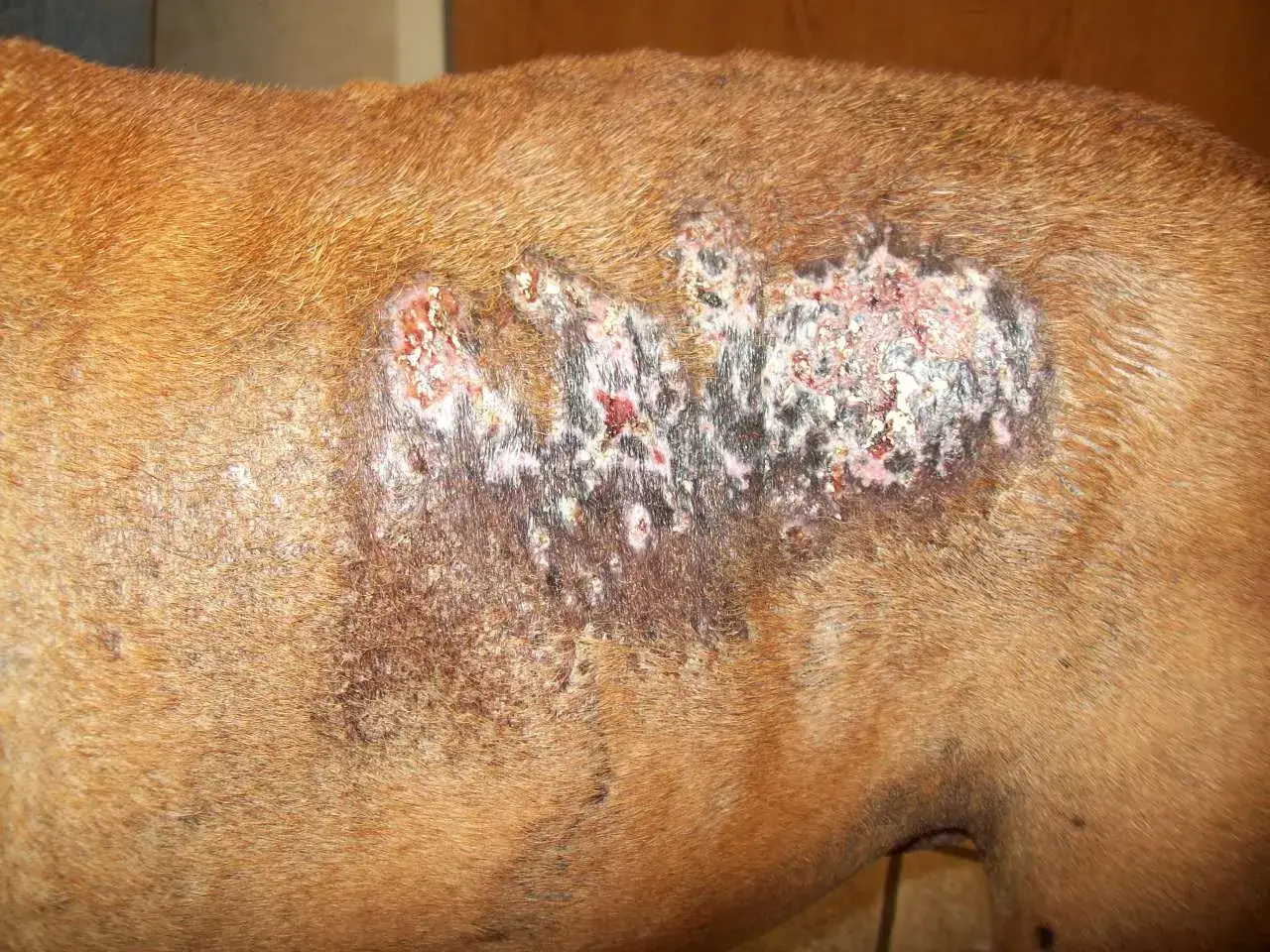
How the lesions usually look and where they show up
The classic lesions are firm papules or plaques with a gritty surface. They can be pink, white, or gray, and they may stay quiet for a while before becoming red, crusted, itchy, or ulcerated. When the deposits work their way out through the skin, they can leave a chalky, white-to-yellow discharge that is easy to mistake for pus.
Common locations include the groin, armpits, back, neck, ventral abdomen, and sometimes the face. In dogs with Cushing's disease, I also keep an eye out for the broader pattern: hair loss, a pot-bellied abdomen, thin skin, blackheads, muscle loss, and increased drinking or urinating. Those signs matter because they often point to the underlying endocrine problem rather than the skin alone.
The localized form behaves differently. A young, large-breed dog may develop a single hard lump at a pressure point or, less commonly, on the tongue. That pattern is a useful reminder that not every calcium deposit on a dog's skin means the same thing, and the location can give your veterinarian an important clue. From there, the next step is figuring out why the deposits formed.
Why it happens and which dogs are at higher risk
In practice, the most common trigger is too much steroid effect in the body. That can happen because the dog has Cushing's disease, because a steroid medication has been used for too long, or because both are true at the same time. The skin is very sensitive to that hormonal environment, and mineralization can follow.
Less commonly, calcification in the skin can be part of a broader metabolic or systemic problem. Kidney disease, diabetes, lung disease, and certain cancers can all play a role in some dogs, especially when calcium and phosphate balance is disrupted. Serious illness can also change how the body handles minerals, which is why I never want to treat the skin in isolation if the pattern looks generalized.
Risk is highest in dogs that have been on long-term corticosteroids or that show other signs of hyperadrenocorticism. For the localized, circumscribed form, I think more about young, large-breed dogs and sites of repeated friction or prior injury. That difference is easy to miss if you only look at the bumps, so the medication history and the dog's age really matter. Once risk is clear, the diagnosis becomes much more targeted.
How veterinarians confirm the diagnosis
Diagnosis usually starts with a good history and a hands-on skin exam. I would want to know whether the dog is taking steroids, how long the lesions have been present, whether they itch or leak, and whether there are signs that suggest Cushing's disease or another internal disorder. Sometimes the skin problem shows up before the more obvious endocrine signs, so a careful exam matters.
The core tests often include bloodwork and urinalysis to look for clues about the underlying disease. If Cushing's is suspected, your veterinarian may recommend hormone testing such as an ACTH stimulation test, a low-dose dexamethasone suppression test, or a urine cortisol-to-creatinine ratio, sometimes along with abdominal imaging. Those tests do not diagnose the skin condition itself, but they help identify what is driving it.
A skin biopsy is usually what confirms the diagnosis. That may sound more involved than it is: the sample is examined under the microscope to prove that calcium is actually present in the skin and to rule out look-alike problems. I value that step because crusted plaques, nodules, and draining lesions can mimic infection, tumors, or other inflammatory skin diseases. Once the diagnosis is confirmed, treatment can be aimed where it belongs.
Treatment is about controlling the cause first
This is the point where many owners hope for a quick cream or a simple pill, but calcinosis cutis rarely works that way. The underlying cause has to be controlled first, or the calcium deposits tend to keep forming.
| Approach | When it helps | What to expect |
|---|---|---|
| Gradual steroid taper or medication change | Dog is on long-term glucocorticoids | Must be done slowly and under veterinary guidance; this is often the most important step |
| Treatment for Cushing's disease | Hormone testing confirms hyperadrenocorticism | Skin changes usually improve only after cortisol control improves |
| Topical DMSO | Localized plaques or persistent mineralized areas | Used off label in dogs; response varies, but it may help soften deposits and support reabsorption |
| Antibiotics and wound care | Ulcerated, inflamed, or infected lesions | Helps secondary pyoderma and discomfort, but does not remove the root cause |
| Surgery | Single, isolated calcinosis circumscripta lesion | Can be curative when the lesion is localized and the trigger is removed |
I am careful not to promise fast cosmetic improvement. Even after the trigger is controlled, the deposits can take months to resolve, and some lesions look worse before they look better because the calcium works its way outward through the skin. If there is a secondary infection, that needs treatment too, because a crusted or oozing plaque is much harder for the skin to heal on its own.
One practical rule I always stress: never stop steroids abruptly. If a dog has been on prednisone or another glucocorticoid for a while, the taper has to be planned. That is especially important because the drug may have been prescribed for a serious allergy, autoimmune, or inflammatory condition, and the replacement plan needs to be safe as well as effective. From there, the next concern is what recovery really looks like.
What recovery looks like and how to prevent a repeat
Recovery depends far more on the underlying disease than on the calcium deposits alone. If the trigger is controlled well, the skin may gradually clear, but I would still expect a slow timeline rather than an overnight fix. In dogs with a single localized circumscribed lesion, surgical removal can be very successful and recurrence is uncommon when the lesion was truly isolated.
For dogs that need long-term corticosteroids, prevention is mostly about monitoring and restraint. If a dog seems to need steroids for more than 3 to 4 months, the plan should be re-evaluated. Many dogs on chronic steroids also need regular rechecks, and quarterly exams with blood tests and urine cultures every 6 months are a sensible benchmark to keep complications from sneaking up.
At home, I would watch for new crusty patches, pain, swelling, foul odor, licking, or discharge. Keeping the skin clean and dry, preventing licking when needed, and reporting new lesions early makes a real difference. More than anything, the goal is to catch the problem while it is still a skin clue rather than letting it become a full endocrine or infection problem.
The details that change the outcome most
What matters most is not the calcium itself but the reason it appeared. If the lesion is tied to Cushing's disease or chronic steroid exposure, the best results come from treating that issue directly and then giving the skin time to settle.
When I look at a dog with firm, gritty plaques, I ask three questions first: Is the dog on steroids? Are there signs of Cushing's? Does the lesion look localized or widespread? Those three answers usually point me toward the right path much faster than the skin appearance alone.
If you remember only one thing, make it this: calcinosis cutis is often a warning light, not the engine problem. Once the cause is identified and managed carefully, most dogs have a much better chance of healing comfortably and avoiding repeat flare-ups.